By Defne
The story of how Rosalind Franklin’s work on DNA was used by Watson and Crick to make the first DNA model and how she was unrepresented for her work has become widely known. But what is the full story behind this?
“Rosy Franklin” as presented by James Watson
In his 1968 book, The Double Helix, James Watson gives an autobiographical story of the discovery of DNA’s structure. Despite how central her role was in the discoveries made, the first mention of Rosalind Franklin gives us the impression that Franklin was more of a hindrance to research progress: Watson writes, “it was increasingly difficult to take Maurice’s mind off his assistant, Rosalind Franklin.” I would like to add that Franklin was Maurice Wilkins’ colleague, not assistant. “Clearly,” he goes on to write, “Rosy had to go or be put in her place. The former was obviously preferable because, given her belligerent moods, it would be very difficult for Maurice to maintain a dominant position that would allow him to think unhindered about DNA.”
Just by reading this book, it would be difficult to understand how important Franklin’s contributions were to the breakthroughs regarding DNA.
So let us look at the story from a different angle.
Who actually is Rosalind Franklin?
Rosalind Franklin was a British chemist and biophysicist who was working as a research associate in King’s College, alongside Maurice Wilkins, during the race for the discovery of the DNA (deoxyribonucleic acid) molecules’s structure, where she mostly worked with the X-ray crystallography technique she learnt in Paris.
Franklin’s Discovery
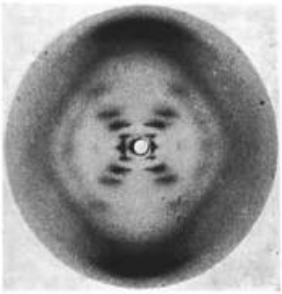
Franklin is famous for the X-ray crystallography image produced by her student Raymond Gosling (see photo 51), the first image clearly showing the double helix structure of DNA. The X-ray crystallography also provided the numbers that were needed to mathematically deduce the full structure of
the molecule.

Photo 51: The X-ray diffraction pattern of DNA (Gosling)
As Franklin was busy with the calculations, she shared some of her data with Max Perutz. At the time, Watson and Crick were working on DNA, trying to deduce the structure before Linus Pauling. Perutz ended up sharing the data with them, from which they based their calculations (without the permission from Franklin) to come up with their famous model before Franklin had finished interpreting it. Her notes show that she ended up finding the correct structure for DNA, but Watson and Crick had already finished their model. (Cobb)

Franklin’s lab notebook, showing her explorations of DNA structure (Wellcome Collection)
Rosalind Franklin died of ovarian cancer in 1958. James Watson, Francis Crick and Maurice Wilkins went on to receive the Nobel Prize for Medicine in 1962 , although at the time Nobel Prizes were still awarded posthumously .
Francis Crick’s 1974 The Double Helix: A Personal View shows a more unbiased view of Franklin’s contribution, in comparison to Watson. When speculating about who would have discovered the structure if not for them, he states: “Rosalind Franklin was only two steps away from the solution. She needed to realise the two bases run in opposite directions and that the bases were paired together.”
Building your own DNA model out of candy
What is the structure actually like?

The picture shows phosphate groups in yellow, 5-carbon deoxyribose sugar in pink and the nitrogenous bases (A, T, G, C) in cyan. There are covalent bonds between the phosphate and sugars, as well as the sugars and nitrogenous bases. DNA has two antiparallel strands, meaning the 5-carbon sugar is facing different directions on the two strands that are attached.
DNA structure (Clark)
For the nitrogenous bases, A is always paired with T via 2 hydrogen bonds and G is always paired with C via 3 hydrogen bonds.
The Craft
With a deeper understanding of the history behind the famous DNA model and how this molecule is structured, it could be fun to build your own model. Since Halloween is nearing, what better material to use for this than candy?
Here’s what you’ll need:
- Candy (I used fruit gums, strawberry laces and starburst)
- Toothpicks
Step 1: Choose a string-like candy to represent your covalent bonds. Lay two of them side by side.
Step 2: Choose a candy for the deoxyribose (I used red fruit gums which are symmetrical, but if you have something asymmetrical you can also show the antiparallel strands!) and phosphates.
Step 3: Choose different colours of the same candy to represent each nitrogenous base (starburst is great for this).
Step 4: Attach the phosphates representations on top of the laces, and deoxyribose underneath. The bases go sideways of deoxyribose. This is one nucleotide. Repeat this process downwards until you are done with the lace. Repeat for the second one and attach the laces from the complementary nucleotides.

Step 5: Twist to get your double helix!
Sources
Clark, Jim. “DNA – Structure.” Chemguide.co.uk, May 2016, http://www.chemguide.co.uk/organicprops/aminoacids/dna1.html.
Cobb, Matthew. “Sexism in Science: Did Watson and Crick Really Steal Rosalind Franklin’s Data?” The Guardian, The Guardian, 14 Feb. 2018, http://www.theguardian.com/science/2015/jun/23/sexism-in-science-did-watson-and-crick-really-steal-rosalind-franklins-data.
Pietzsch, Joachim. “The Nobel Prize in Physiology or Medicine 1962.” NobelPrize.org, http://www.nobelprize.org/prizes/medicine/1962/speedread/.
The Nobel Prize. “Nobel Prize Facts.” NobelPrize.org, 2019, http://www.nobelprize.org/prizes/facts/nobel-prize-facts/.
Watson, James. THE DOUBLE HELIX a Personal Account of the Discovery of the Structure of DNA. 1968, sites.bu.edu/manove-ec101/files/2017/09/Watson_The_Double_Helix.pdf.Wellcome Collection. Reports and Working Notes on DNA. Reference: FRKN 1/4. wellcomecollection.org/works/x4c2x8rr/items?canvas=8.

Leave a comment